Jovana Drinjakovic
Study reveals unexpected diversity in the ways proteins bind to their partners, with implications for understanding the molecular roots of disease.
University of Toronto scientists have developed a new technology to find the exact regions in protein molecules that allow them to bind to other proteins in cells. The finding will help scientists understand how genetic mutations change protein binding to spur disease.
A team led by Professor Sachdev Sidhu, of U of T’s Donnelly Centre for Cellular and Biomolecular Research and Department of Molecular Genetics, focused on a large group of human proteins that control cell growth and are often mutated in cancer. All of these proteins use a part called the SH3 domain to connect with other proteins. The researchers found that the SH3 domains have the potential to bind to a far more diverse set of partners than previously thought, vastly expanding their possible biological roles. The team published its results September 7 in the journal Structure.
Most diseases occur when proteins go awry by, for example, losing the ability to bind to their partners and/or to acquire new alliances.
“Despite being one of the best-studied protein families, our results reveal entirely new and unexpected partnerships and functions for many family members,” says Sidhu, who is also Director of the Centre for Commercialization of Antibodies and Biologics and Senior Investigator at the Ontario Institute for Cancer Research. “In a broader sense, the findings highlight how much still remains to be learned about the basic processes within human cells”.
The study was made possible by a new technology that couples state-of-the-art protein engineering with rapid DNA sequencing, allowing the team to test a staggering 10 billion protein interactions—10 million times more than what is possible with other available methods.
"Our results reveal entirely new and unexpected partnerships and functions for many SH3 domain family members"—Professor Sachdev Sidhu
Proteins are the products of genes that make up our cells and do most of the work in them. By binding to each other, proteins carry out cellular processes which ensure that cells grow and divide in a tightly controlled fashion.
“If you understand molecular rules governing protein interactions, then you can try to predict binding partners, something that’s still very hard to do,” says Joan Teyra, a research associate in the Sidhu lab who worked on the study.
Proteins are made from amino-acids stitched together in a string which then gets folded origami-style to form a three-dimensional protein molecule. Among the folds are the binding sites with which the proteins contact one another. These sites harbor short amino acid patterns that can only be “read” by the correct partners, ensuring that the binding is precise and tight.
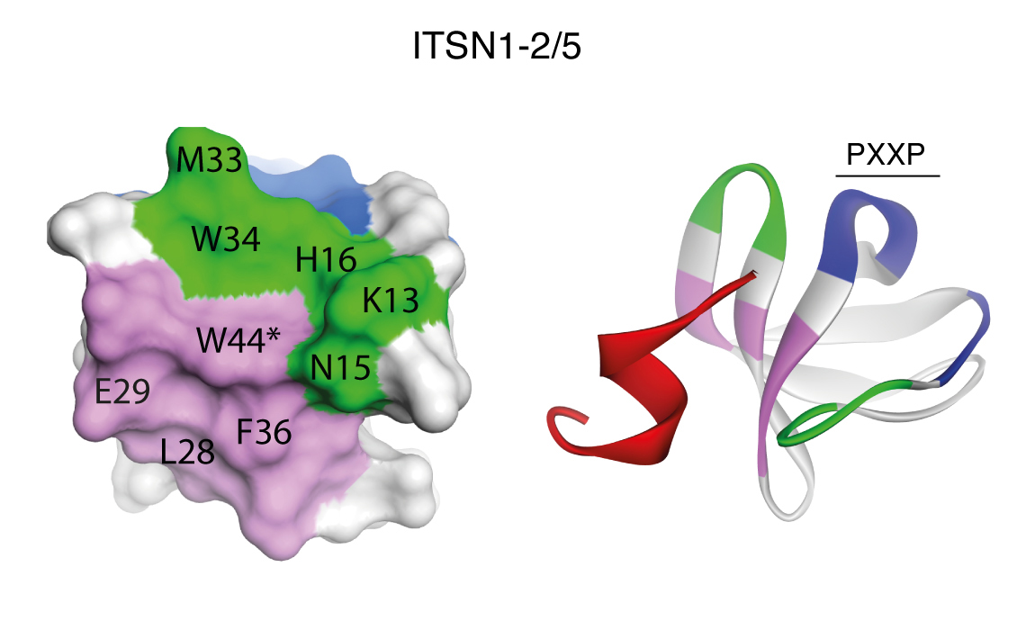
The hunt for the SH3 domains’ interacting partners began in the 1990s when researchers discovered that they bound proteins containing the PxxP pattern, where “P” stands for the amino acid proline and “x” for any amino acid. But the pattern turned out to be too common, found all over the proteins, even in places where binding does not happen.
Teyra and colleagues identified more than 100 new amino acid patterns, in the vicinity of the PxxP, that are key for binding. What’s more, the team found that SH3 domains can bind to large swaths of targets that don’t even contain the PxxP, upending the widely held belief that the PxxP solely dictates the binding.
To do this, the team collaborated with computational biologist Gary Bader, also in the Donnelly Centre, to engineer viral particles that display on their surface protein fragments 12 amino-acids long, covering all possible combinations of the 20 amino-acids found in proteins. The total of 10 billion protein fragments were then doused with 115 different SH3 domains to find the ones that bound tightly, followed by data analysis to identify the exact amino-acid segments that made the binding possible.
“After 30 years of research, this is the most comprehensive mapping study for the SH3 domains,” says Teyra. “We confirmed that PxxP is the most common way of recognizing proteins, but we found that 25 per cent of target fragments don’t even have the PxxP. This is very interesting because it suggests that the SH3 proteins could also be binding totally different proteins from what we are used to and that vastly expands the biology field.”
This means that the researchers can now cast a wider net in search of proteins capable of binding the SH3 domain to reveal new ways in which these proteins work. The key to this follow up research will be to integrate data from other fields and studies, said Teyra.
The research was supported by the funding from the Canadian Institutes of Health Research.